Unit 3: The CellThe cell is the most basic or fundamental unit of life, the smallest thing we can observe that still retains the characteristics of life discussed in our first unit. In this unit we will explore the structure of cells to gain an understanding of their enormous complexity and responsiveness to their environment. We will begin by defining the basic characteristics all cell shared the two basic categories all cells fit into before expanding on some of the more complex peculiarities we can observe in specific cell types. As we progress through the unit we will compare animal and plant cells identifying and describing the variety of different organelles we can observe in these cells and what role they play in supporting the cell as a whole. We will study the endosymbiotic theory to see how cells have evolved over time and take an introductory look at basic taxonomy. And finally, we will study stem cells to better understand their significance to organisms and emerging medical technologies.
|
|
What is a Cell?
"All life is made of cells and the cell is the most basic unit of life." This should sound pretty familiar, you learned this back in Unit 1 when we were discussing the characteristics of life. In fact, these concepts come from what is known as the Cell Theory which states:
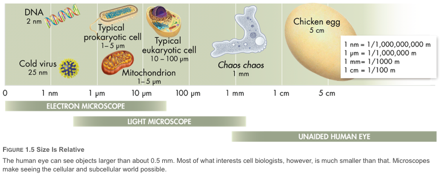
Cells are very small and generally cannot be seen by the unaided human eye. It is no surprise then that cells were not discovered until the 1600s, when microscopes became strong enough to see them. In 1665, Robert Hooke, placed a small sample of cork under a compound light microscope and saw what he described as "small chambers". Hooke settled on calling these chambers "cells" because they reminded him of the rooms or cells in a monastery. Around the same time in Holland, Anton van Leeuwenhoek observed pond water under a light microscope and observed what we know today as bacteria. Soon scientists realized that all life was made of cells.
While the idea of what a cell is may seem straight-forward, cells themselves are anything but simple. They are incredibly diverse and complex in terms of their size, function and design and yet all cells do share certain key traits:
- All living things are made up of cells.
- Cells are the most basic unit of structure and function in living things.
- All cells come from pre-existing cells.
Cells are very small and generally cannot be seen by the unaided human eye. It is no surprise then that cells were not discovered until the 1600s, when microscopes became strong enough to see them. In 1665, Robert Hooke, placed a small sample of cork under a compound light microscope and saw what he described as "small chambers". Hooke settled on calling these chambers "cells" because they reminded him of the rooms or cells in a monastery. Around the same time in Holland, Anton van Leeuwenhoek observed pond water under a light microscope and observed what we know today as bacteria. Soon scientists realized that all life was made of cells.
While the idea of what a cell is may seem straight-forward, cells themselves are anything but simple. They are incredibly diverse and complex in terms of their size, function and design and yet all cells do share certain key traits:
- All cells have a cell membrane to separate their contents from the environment and help to establish homeostasis.
- All cells contain genetic material in the form of DNA.
- All cells contain small structures called organelles that each have specific functions and work to support the cell as a whole.
Prokaryotic vs. Eukaryotic Cells
|
Prokaryotic Cell
|
Eukaryotic Cell
|
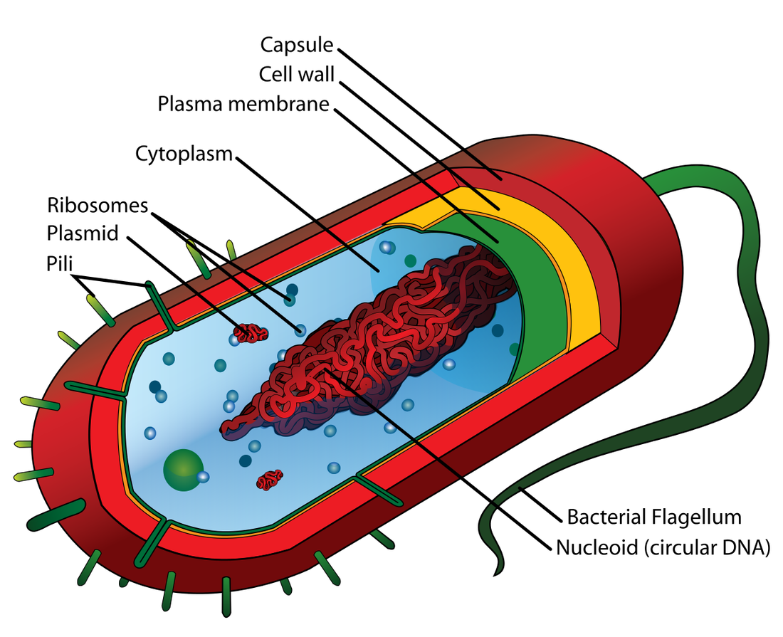
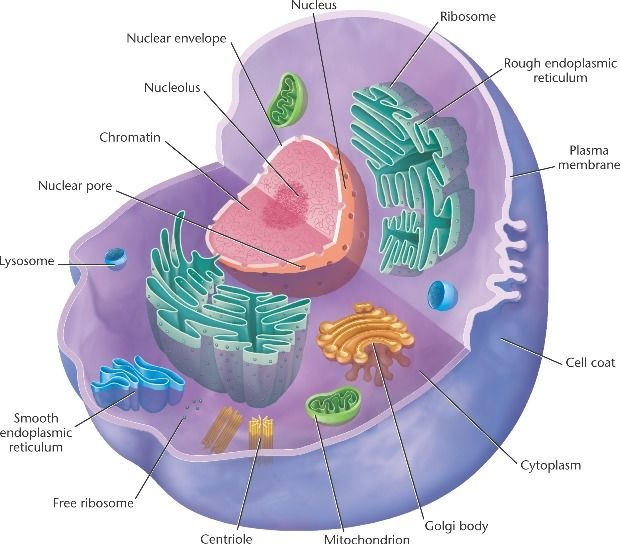
Prokaryotic cells (also known as prokaryotes) were the first type of cell to appear on Earth about 3.8 billion years ago. Prokaryotic cells tend to be smaller and simpler than eukaryotic cells, although this is not always the case. The primary difference between a prokaryotic cell and a eukaryotic cells is that prokaryotes do not separate their genetic material from the rest of the cell. In contrast, eukaryotic cells (also called eukaryotes) have a special organelle called the nucleus to house and protect the DNA of the cell. The nucleus is composed of a nuclear envelope that surrounds the DNA and contains small pores that allow the passage of RNA molecules during proteins synthesis (more on that later).
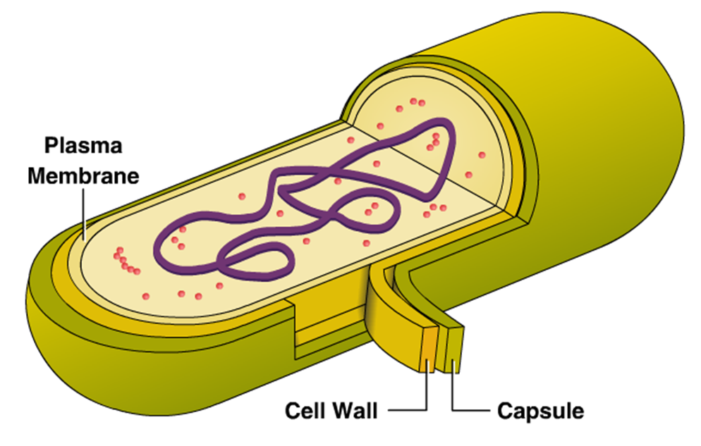
Another difference between prokaryotic and eukaryotic cells is that eukaryotes have a greater diversity of organelles available to them and organelles are typically surrounded by a protective membrane. On the other hand, prokaryotic organelles are not membrane bound. Finally, prokaryotes are generally unicellular and function independently, while eukaryotes are generally multicellular and their cells express a high degree of interdependence on one another. Because eukaryotic cells rely so much on one another, they have special forms of communication and lack higher levels of cellular protection, such as a capsule, seen in prokaryotes.
Prokaryotes vs. Eukaryotes Summarized
- Prokaryotes lack a nucleus, but eukaryotic cells separate their DNA from the rest of the cell using a nucleus.
- Prokaryotic cells tend to be smaller than eurkaryotic cells; on the order of about 10-20x smaller.
- Eukaryotic cells have a wider variety of organelles that prokaryotic organelles, including endoplasmic reticulum (smooth and rough), Golgi apparatus and mitochondria.
- Prokaryotic cells tend to be unicellular, where as eukaryotic cells are usually multicellular.
- Prokryotic cells are generally independent and can operate/survive on their own where as eukaryotic cells depend on other cells (from the same orrganism) for survival.
|
Cell Type and Taxonomy
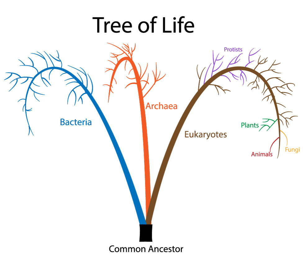
Taxonomy is the science of classifying organisms according to the evolutionary history and anatomical traits. Organisms are placed into different categories depending on how closely related they are. These categories include domain, kingdom, phylum, class, order, family, genus and species (from broadest to most specific). Under current taxonomy, living organisms can be divided up into 3 domains, the highest level of organization: Archaea, Bacteria and Eukarya. Archaea are a very ancient type of life form that tend to live in extreme environments: halophytes ("salt lovers"), thermophiles ("heat lovers"), acidophiles ("acid lovers") etc. This has earned the Archaea the nickname of extremophiles. In comparison, Bacteria are the most prolific organisms on Earth with about 40,000,000 bacteria cells in a single gram of soil. |
|
In fact, bacteria cells outnumber the human cells in your body by about 10:1. Bacteria play a number of important ecological roles in ecosystems including decomposition of dead organisms and recycling of nutrients like nitrogen. The organisms grouped into domains Bacteria and Archaea are prokaryotic and species under the Eukarya domain are eukaryotic and include the plant, animal, fungi and protist kingdoms. This means that cell type (prokaryotic or eukaryotic) is one of the most important traits in taxonomy.
Animal vs. Plant Cells
|
|
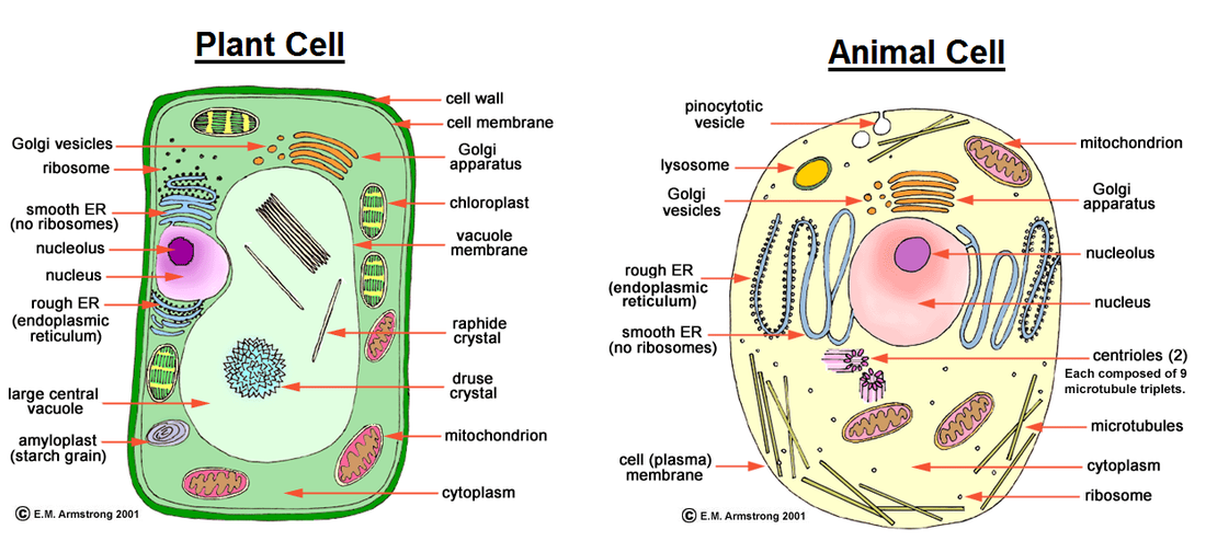
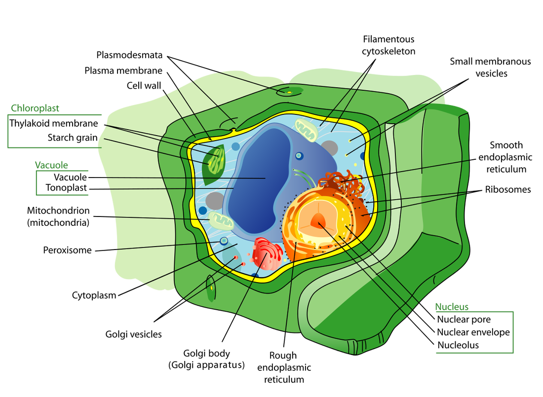
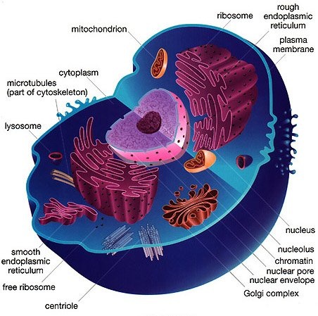
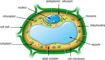
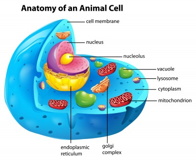
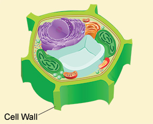
Just as we can make certain generalities between the prokaryotes and eukaryotes, we can make important distinctions between animal and plant cells. The surface of an animal cell is typically rounded and smooth because animal cells are only surrounded by a cell membrane. Plant cells however, tend to have straight edges due to the presence of a thick, reinforced cell wall that provides additional rigidity and strength to the cell. Why do plant cells have this cell wall, while animal cells do not? Plants are immobile and lack a skeleton; they use their cell walls to provide them with support to stand up straight. Remember, plants are competing for sunlight so the taller they are, the more well off they will be. In contrast, most animals are highly mobile, requiring a high degree of flexibility. Cell membranes are very flexible, but cell walls would prevent animals from moving round and acquiring food and other resources.
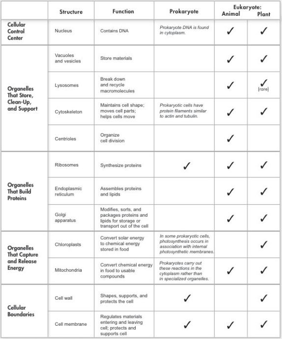
There are of course other important differences between animal and plant cells. For one, plant cells contain a chloroplast, allowing them to undergo photosynthesis, while animal cells lack this organelle entirely. This is because plants are autotrophs and must produce their own organic food sources from inorganic compounds, while animals are heterotrophs and consume tissues for energy. In addition, plant cells generally have a single, massive vacuole to store water, while animal cells have multiple smaller vacuoles. The reason for this is that plant cells use their vacuole to generate turgor pressure (internal water pressure) and keep the plant upright. Finally, animal cells contain centrioles that help in organizing cellular division, but plant cells lack these organelles. The table below summarizes the similarities and differences between prokaryotic, animal and plant cells.
Animal vs. Plant Cell Summary
- Animal cells are typically round and smooth, while plant cells tend to have straighter edges and appear more cubical or boxy due to cell walls.
- Animal cells lack cell walls and only have a cell membrane. Plant cells have both a cell wall and cell membrane. The cell wall in plant cells have have varying degrees of thickness depending on the specific plant cell type.
- Animal cells lack the chloroplasts found in plant cells, but do have centrioles to aid in cellular division. Plant cell division is slightly different from animal cell division and hence lacks centrioles.
- Animal cells have multiple, smaller vacuoles to stores nutrients, while plant cells have one large, central vacuole that is primarily used to store water and provide turgor pressure and maintain the shape of the plant cell. In a plant cell, you can think of the vacuole as a swollen water balloon in a shoe box. The water balloon pushes outward and helps keep the shoebox rigid.
The Organelles
|
Organelles literally translates to "little organs". Organelles are the small subunits of cells that help the cell function and survive. Each organelle has an important function. There are a great number and variety of different organelles, ranging from the DNA storing nucleus, to the protein-producing ribosome. The key to understanding and remembering them all will be your ability to organize the organelles in a meaningful way. I have done my best to organize the organelles in a cohesive, intuitive manner, but this organization may be the best one for you. The key will be finding whatever way helps you to remember the function of these organelles.
|
|
Structural Organelles
|
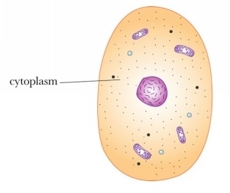
Cytosol and Cytoplasm
The cytosol is the gel-like matrix that fills the inside of all cell and does not include the organelles within the cell. The cytosol exists within the cell membrane, but outside of the nucleus (prokaryotic cells also have cytoplasm despite lacking a nucleus). The term cytoplasm refers to the cytosol with the organelles suspended in it. The cytosol holds and supports the other organelles with in the cell. The cytosol is mostly composed of water, salt and various proteins and would test positively with Biuret's reagent because it contains proteins. |
|
|
Cytoskeleton
The cytoskeleton is literally the backbone of the cell (cyto = cell and skeleton = ...well skeleton) and is present in all cell types. In addition to holding the cell up and giving it shape, the cytoskeleton provides a road network for moving substances throughout the cell. The cytoskeleton can even help the cell move when extended out of the cell as flagella and cilia. The cytoskeleton is composed of microfilaments and microtubules and found in all cell types. Microfilaments are composed of proteins called actin and help in cellular movement. Microtubules are composed of proteins called tubulins and are move involved with cell shape. Microtubules also play a key role in cellular division as the mitotic spindle. During mitosis, the centrioles form the spindle fibers that help to separate the homologous chromosomes (more on that later). Because the cytoskeleton is composed of microfilaments and microtubules, which are in turn composed of different proteins (actin and tubulins respectively), the cytoskeleton would test postively when tested with biuret's reagent. |
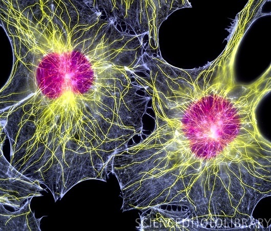
In the graphic above, the cytoskeleton is highlighted with the cell using fluorescent taggers.
|
Centrioles
Like the cytoskeleton, the centrioles are formed of microtubules. This organelle helps to organize cellular division in animal, but not plant cells, by producing the spindle fibers. Again, the centrioles would test positively when tested with biuret's reagenet because they are composed of microtubules which are made of the protein tubulin.
Like the cytoskeleton, the centrioles are formed of microtubules. This organelle helps to organize cellular division in animal, but not plant cells, by producing the spindle fibers. Again, the centrioles would test positively when tested with biuret's reagenet because they are composed of microtubules which are made of the protein tubulin.
|
Spindle Fibers
In animal cells, the spindle fibers are produced by the centrioles and help to move and separate the chromosomes during cellular division. The spindle fibers are composed of microtubules that can quickly be built and disassembled. Each spindle fiber is composed of a single microtubule and hence would test positively when tested with biuret's reagent. |
|
Economic Organelles
|
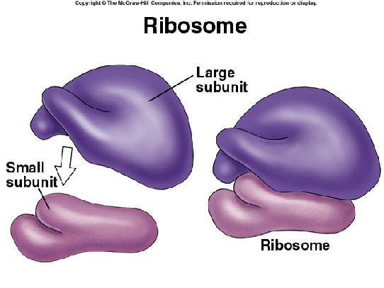
Ribosomes
Ribosomes are relatively small organelles found in all cell types, built out of RNA (rRNA) and a variety of proteins. Each ribosome has two parts: a large and small subunit. These subunits combine together to translate an mRNA transcript (built using DNA as a template during transcription...more on that later) into protein. Ribosomes can be found freely in the cytoplasm or attached to the endoplasmic reticulum (this forms the rough ER). The ribosomes primary goal is to produce new proteins for the cell. We will investigate how ribosomes operate in greater detail when we explore protein synthesis in a later unit. Ribosomes are found in all cell types. Based on its structure, a ribosome would return a positive result when tested with biuret's reagent. |
|
|
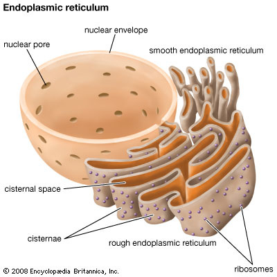
Endoplasmic Reticulum (ER)
The Endoplasmic Reticulum (ER) is an internal membrane system found within eukaryotic cells around the nucleus. This is where lipid and proteins molecules are assembled and packaged. The ER formed from the infolding of the cell membrane, which explains its similar structure composition to the cell membrane, nuclear envelope (of the nucleus), Golgi Apparatus and vesicles. All 5 of these organelle types are composed of a phospholipid bilayer and would return a postive result when tested with Sudan test. The rough ER earns is named because it is lined with ribosomes, making it appear rough in and texture and is found in cells that produce large quantities of proteins using the ribosomes. Some proteins may still be formed on free ribosomes outside the rough ER. Before the proteins are released from the rough ER, they are packaged into vesicles that bud off the membranous rough ER. The smooth ER lacks the ribosomes found on the rough ER (hence the name) and is responsible for building and packaging lipid molecules. Just as in the rough ER, lipid molecules are packaged into vesicles formed from the smooth ER itself. Often the smooth ER contains enzymes to help build these lipid molecules. |
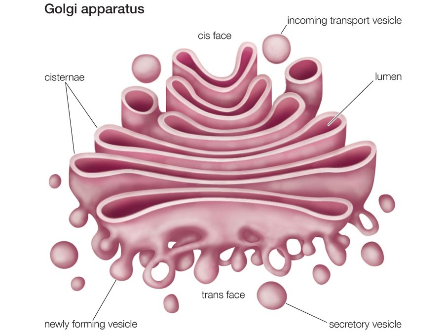
Golgi Apparatus
The Golgi Apparatus is another organelle found only in eukaryotes and resembles a flattened stack of membranes, similar to the ER. Like the ER, the Golgi Apparatus formed from the infolding of the cell membrane. This means the Golgi Apparatus shares the same lipid bilayer structure as the cell membrane, ER and vesicles. Here proteins are collected and modified from the rough ER and shipped to their final destination again by vesicle transport. Again, based on its structure, the Golgi apparatus would test positive for lipids using the Sudan test.
The Golgi Apparatus is another organelle found only in eukaryotes and resembles a flattened stack of membranes, similar to the ER. Like the ER, the Golgi Apparatus formed from the infolding of the cell membrane. This means the Golgi Apparatus shares the same lipid bilayer structure as the cell membrane, ER and vesicles. Here proteins are collected and modified from the rough ER and shipped to their final destination again by vesicle transport. Again, based on its structure, the Golgi apparatus would test positive for lipids using the Sudan test.
|
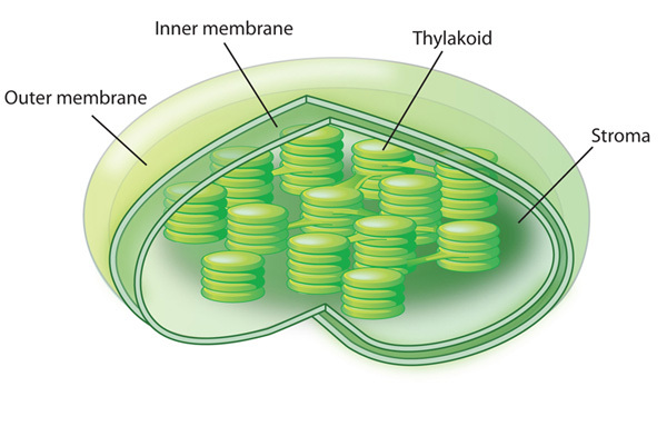
Chloroplast
The chloroplast is a double bound organelle present in plant cells. The chloroplast is the organelle where photosynthesis occurs (photosynthetic prokaryotes lack this organelle and photosynthesis occurs in the cytoplasm) and is primarily responsible for converting carbon dioxide, water and solar energy into simple sugars such as glucose. Chloroplasts contain the pigment chlorophyll which allows them to absorb solar energy and gives them their green color. As you can see in the diagram, the chloroplast has two membranes and contains thylakoid discs suspended in a liquid medium called the stroma. Later, we will see how these contribute to photosynthesis. Be sure to read "Endosymbiotic Theory" below to learn more about chloroplasts and mitochondria. |
|
|
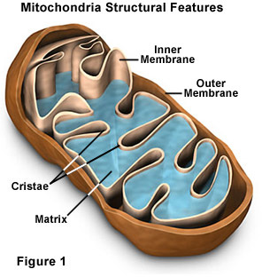
Mitochondria
The powerhouse of the cell- that's how most people refer to the mitochondria. Mitochondria are highly complex and important organelles that convert energy stored in macromolecules (usually carbohydrates) into usable energy as ATP found in all eukaryotic cells. Like chloroplasts, mitochondria are relatively large, have two membranes and are are present in nearly all eukaryotic cells. We will investigate the structure and function of the mitochondria in a later unit on cellular respiration. Because aerobic cellular respiration is the most efficient type of cell respiration, mitochondria need access to plenty of oxygen gas. Mitochondria are found in eukaryotic cells. Be sure to read "Endosymbiotic Theory" below to learn more about chloroplasts and mitochondria. |
|
|
Vesicles
Vesicles are membrane bound sacs that help to transport large quantities of molecules around the cell in eukaryotesand are the primary mechanism of endocytosis and exocytosis (see below). Vesicles share the same lipid bilayer seen in cellular membranes, the ER and Golgi Apparatus and can be formed by the budding (pinching off) of these organelles. This also allows vesicles to easily fuse with the plasma membrane, ER and Golgi Apparatus to export and/or deliver molecules. Based on their sturcture, vesicles would test positive for lipids using the Sudan test because of their phospholipid bilayer. The modified vesicles (vacuoles, lysosomes and peroxisomes described below) would also test positively for lipids for this same reason. |
|
|
Vacuoles
Vacuoles are modified vesicles that store excess nutrients in eukaryotes. Like vesicles and the cellular membrane, vacuoles have a lipid bilayer surrounding them meaning they will test positively for Sudan test based on their structure. Plant cells typically have one large, central vacuole that fills with water which helps to keep plants rigid by generating turgor pressure. When the vacuole is full and swollen with water, the cell is said to be turgid and the plant remains upright. The cell wall prevents the plant cell from bursting. As water is drained from the vacuole, the turgor pressure of the cell decreases and the plant begins to wilt. If the vacuole empties completely, the cell is said to plasmolyze and the cell membrane collapses inwards. Animal cells have multiple smaller vacuoles for storing nutrient resources. Vacuoles are not used to generate turgor pressure in animal cells and take up far less space. |
|
|
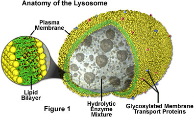
Lysosomes
Lysosomes are specialized vesicles that contain digestive enzymes in animal cells and more rarely, in some plant cells. Lysosomes function like a "clean up" crew or a recycling center that digests excess nutrients (carbohydrates, lipids and proteins) and break down old organelles for reuse within the cell. Lysosomes are typically found in animal cells, but some plant cells will also contain lysosomes. In addition to testing positively under the Sudan test, lysosomes would also test positively under biuret's reagent because they contain digestive enzymes (remember, enzymes are proteins). |
Defensive Organelles
|
Nucleus
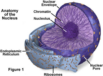
The nucleus protects and houses the DNA in eukaryotic cells. There is only one nucleus per cell (an exception being muscle cells) Considering that DNA contains all the genetic information for the cell and the instructions for building all the proteins needed for cellular activity, the nucleus is often considered the command center of the cell. The nuclear envelope, composed of two phospholipid bilayers, surrounds the nucleus and each membrane has a similar structure to the plasma membrane that surround the cell. Like the ER and Golgi apparatus, the nucleus originally formed from the infolding of the cell membrane. |
|
The nuclear envelope is largely impermeable and strictly regulates what can enter the nucleus, thereby protecting the DNA. The nuclear envelope contains small openings called nuclear pores where mRNA and ribosomes can exit the nucleus (you will learn more about that later in the lear when we study protein synthesis). Because of its shared structure (again similar to the ER, Golgi apparatus, vesicles and cell membrane), the nuclear envelope would test positively for lipids using Sudan test. Within the nuclear envelope lies the chromatin, the loosely packaged, thread-like mass of DNA. In the chromatin, DNA is wrapped around specialized proteins called histones, helping to condense the DNA and package it into the nucleus. Based on its structure, the chromatin would test positively when tested with biuret's reagent. And finally, the nucleolus is a dense region with in the nucleus where ribosomes are formed.
|
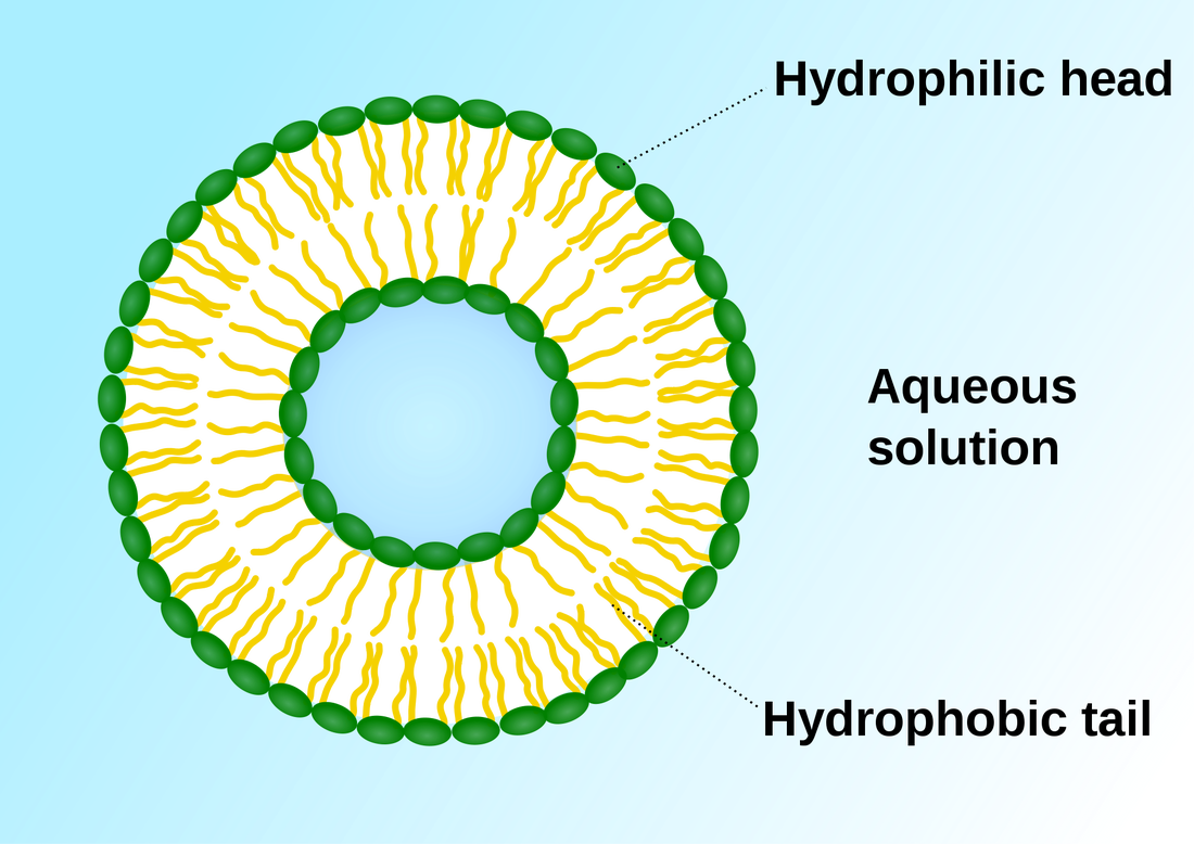
Cell Membrane
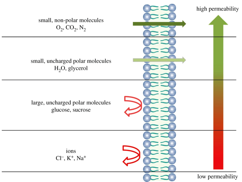
The cell membrane is found in all cell types and surrounding and protecting and cell and separating the cell and its contents from the surrounding environment. As you know, the cell membrane (also called the plasma membrane) is composed of phospholipids, more specifically a phospholipid bilayer. The plasma membrane is selectively permeable which means only certain atoms and molecules can pass through it into the cell. This protects the cell from dangerous toxins and compounds that can be found in the extracellular fluid surrounding the cell. In general, only small, uncharged atoms and molecules may pass through the cell membrane at will. Larger and/or charged/polar molecules or ions cannot pass through the plasma membrane at will. |
|
So the permeability of the cell membrane is determined by two variables: size and electrical charge (including polarity). The reason smaller substances can pass through the cell membrane with ease is because of their size. Smaller substances like oxygen gas can easily squeee between the phospholipids of the cell membrane and enter or exit the cell. Uncharged substances pass through the cell membrane unhindered because they do not interact with the phospholipids. Charged or polar substances interact with the phopholipids (which have a polar head) and can get stuck as thry try to move across the cell membrane.
Water is a small molecule capable of passing through the plasma membrane, but its polarity hinders it greatly and so water relies on other means to enter the cell. The cell membrane's selective permeability can be thought of as a two-edged sword, both helping and harming the cell: while the cell membrane does not allow dangerous toxins into the cell, it can also prevent vital nutrients like potassium ions or glucose from entering the cell.
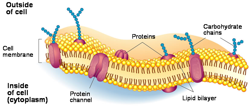
The plasma membrane is often lined with many proteins that help move nutrients into and out of the cell, compensating for its "two-edged sword effect". These processes are highly regulated- only specific molecules react with the membrane proteins and are allowed to pass through the cell membrane. Protein pumps and channels are examples of such proteins. Based on its structure, the cell membrane would return a psotive result when tested with Sudan test and Biuret's reagent. You may notice in the diagram below that there are also carbohydrate chains in the cell membrane, but we will be ignoring these for our class.
Water is a small molecule capable of passing through the plasma membrane, but its polarity hinders it greatly and so water relies on other means to enter the cell. The cell membrane's selective permeability can be thought of as a two-edged sword, both helping and harming the cell: while the cell membrane does not allow dangerous toxins into the cell, it can also prevent vital nutrients like potassium ions or glucose from entering the cell.
The plasma membrane is often lined with many proteins that help move nutrients into and out of the cell, compensating for its "two-edged sword effect". These processes are highly regulated- only specific molecules react with the membrane proteins and are allowed to pass through the cell membrane. Protein pumps and channels are examples of such proteins. Based on its structure, the cell membrane would return a psotive result when tested with Sudan test and Biuret's reagent. You may notice in the diagram below that there are also carbohydrate chains in the cell membrane, but we will be ignoring these for our class.
|
Cell Wall
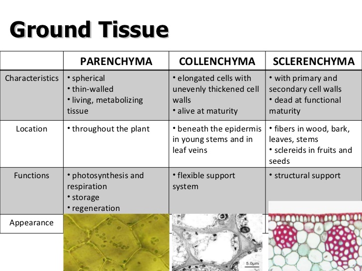
Cell walls are reinforced structures that surround cells. They exist beyond the cell membrane can are composed of different materials depending on the organism: cellulose and sometimes lignin in plants; chitin in fungi; and peptidoglycan in bacteria. These materials contain complex carbohydrates and are indigestible in most animals including humans and provide us with dietary, insoluble fiber. Animal cells lack a cell wall because they rely on movement for survival. Movement requires flexibility and so cell walls would only slow down animals. Plants on the other hand, do not move and lack a skeleton to provide their bodies with support and therefore rely on cell walls to provide rigidity and strength. Like the cell membrane, cell walls are largely impermeable. In plants, cell walls can come in various degrees of thickness. All plant cell cell walls contain cellulose (a complex carbohydrate) with only some containing a molecule called lignin (a complex carbohydrate) which is used to reinforce cells. Parenchyma cells are protected by the thinnest cell walls and compose the "soft" parts of plants like leaves and small stems. Cell walls in collenchyma cells offer a more medium level of protection as seen in the fibers of a stalk of celery. Parenchyma and collenchyma cell cell walls both contain cellulose with no lignin. The major difference between the two is that collenchyma cell cell walls are thicker and therefore contain more cellulose. |
|
Finally sclerenchyma cells often the maximum level of cell wall protection; their cell walls are composed of cellulose and lignin. Sclerenchyma cells are the fundamental component of xylem and phloem and offer enhanced support to the plant, especially trees: the strength of wood comes directly from this type of cell. Sclerenchyma cells actually have two cell walls to further reinforce their strength and rigidity and are dead at maturity.
Having cells of varying cell wall strength allows the plant to be flexible in some locations and strong in others where support is required. Being more flexible can reduce damage from wind or passing animals and being more rigid can help the plant stand up right, especially in large, heavy plants like trees. This is why parenchyma cells are often found in leaves and twigs, collenchyma cells are found in the stems and sclerenchyma cells are found in tree trunks.
You should know that a plant cell wall will always test positively for complex carbonhydrates because of the presence of cellulose and lignin, both of which are polysaccharides. In addition, fungal cell walls will also test positively for Lugol's Iodone test because they contain chitin, a complex carbohydrate. Peptidoglycan contains both carbohydrates and proteins and so a bacteria cell wall would test positively for both Lugol's Iodine and Biuret's reagent tests.
|
Capsule
The capsule is an additional level of cellular protection found in many bacteria cells. The capsule is composed of polysaccharides and helps to increase the virulence (disease causing factor) of the bacteria by making it difficult for phagocytes (white blood cells) to consume and destroy the bacteria. In addition, because the capsule exists around the cell wall of the bacteria it can render antibiotics ineffective as these chemicals target the peptidoglycan structure of the cell wall. Capsules are not found in eukaryotic cells and will test positively under Lugol's Iodine test. |
|
|
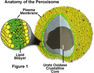
Peroxisome
Like vacuoles and lysosomes, peroxisomes are modified vesicles. Peroxisomes are most closely related to lysosomes as both organelles contain digestive enzymes. However, unlike lysosomes (which primarily act as a recycling center for the cell by breaking down old organelles and macromolecules), peroxisomes use their digestive enzymes to break down dangerous toxins and chemicals in the cell. For example, peroxisomes often contain catalase to help them break down hydrogen peroxide, a dangerous toxin cells produce as a byproduct of normal metabolic activities. A final distinguishing feature of peroxisomes is the presence of a crystalized core in some, but not all peroxisomes. In addition to testing positively under the Sudan test, lysosomes would also test positively under biuret's reagent because they contain digestive enzymes (remember, enzymes are proteins). |
Cellular Exchange Organelles
|
Protein Channels
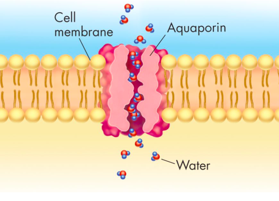
As you know the plasma (cellular) membrane is selectively permeable and only allows certain atoms and molecules into the cell. This protects the cell, but also limits what nutrients can enter/exit the cell. To remedy this, the cell uses protein channels, proteins embedded within the cell membrane that form channels or passages through the cell membrane. Of course just like the cell membrane itself, these protein channels are very selective- they only allow specific molecules to pass through them, all others are excluded. For example, aquaporins (its use is in the name, aqua/water pore in) are protein channels that allow water molecules to enter and exit the cell. Only water may pass through the aquaporin, other molecules and chemicals like glucose may not pass through the aquaporin. Protein channels, including aquaporins, do not require energy (ATP) to function, they are passive and rely on diffusion- the natural process by which molecules spread from areas of high concentration to areas of lower concentration. |
|
Protein Pumps
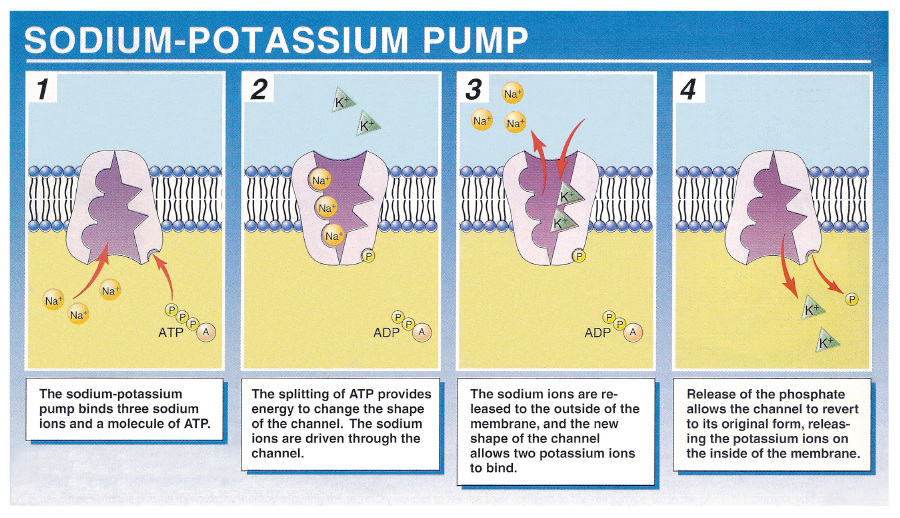
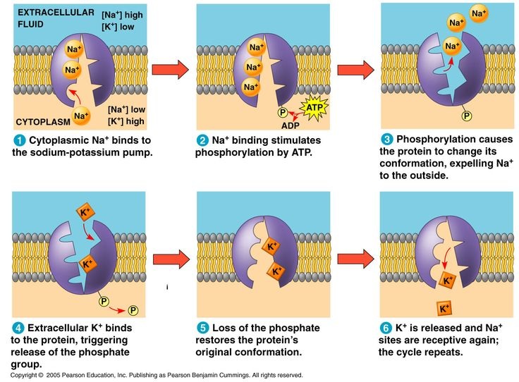
Like protein channels, protein pumps are proteins embedded in the cell membrane that help to transport molecules and ions into and out of the cell. However, unlike protein channels, which are really nothing more than a protein tunnel through cell membrane, protein pumps actively move or pump molecules across the membrane. This requires energy in the form of ATP which is a major drawback of protein pumps compared to protein channels, but one of the major advantages of protein pumps is that they can move molecules or ions against their concentration gradient; from areas of low concentration to high concentration, the reverse of diffusion. An example of a protein pump would be the sodium-potassium pump. In this system, 3 sodium ions from within the cell bind to the pump. Next ATP is applied and the sodium are expelled as the protein pump changes its shape. Finally, two potassium ions bind to the protein pump and are forced into the cell as the protein pump returns to its original shape. This allows the cell to build up large concentrations of vital nurtients inside the cell. The other major advantage to protein pumps is that they can move substances across the cell membrane much more quickly than protein channels can using diffusion.
Like protein channels, protein pumps are proteins embedded in the cell membrane that help to transport molecules and ions into and out of the cell. However, unlike protein channels, which are really nothing more than a protein tunnel through cell membrane, protein pumps actively move or pump molecules across the membrane. This requires energy in the form of ATP which is a major drawback of protein pumps compared to protein channels, but one of the major advantages of protein pumps is that they can move molecules or ions against their concentration gradient; from areas of low concentration to high concentration, the reverse of diffusion. An example of a protein pump would be the sodium-potassium pump. In this system, 3 sodium ions from within the cell bind to the pump. Next ATP is applied and the sodium are expelled as the protein pump changes its shape. Finally, two potassium ions bind to the protein pump and are forced into the cell as the protein pump returns to its original shape. This allows the cell to build up large concentrations of vital nurtients inside the cell. The other major advantage to protein pumps is that they can move substances across the cell membrane much more quickly than protein channels can using diffusion.
|
Plasmodesmata
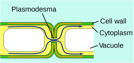
Like protein pumps, plasmodesmata move nutrients and other atoms and molecules in and out the cell using ATP. However, unlike protein pumps which traverse the cell membrane, plasmodesmata (which are only found in plant cells) span across the cell membrane and cell wall. Plasmodesmata enable transportation and communication between cells, which is of course very important in multicellular organisms. In fact, plasmodesmata directly connect the cytoplasm of adjacent cells in plants. The plasmodesmata forms a bridge between the two cells and can be turned on or off by opening and closing the channel using ATP. |
|
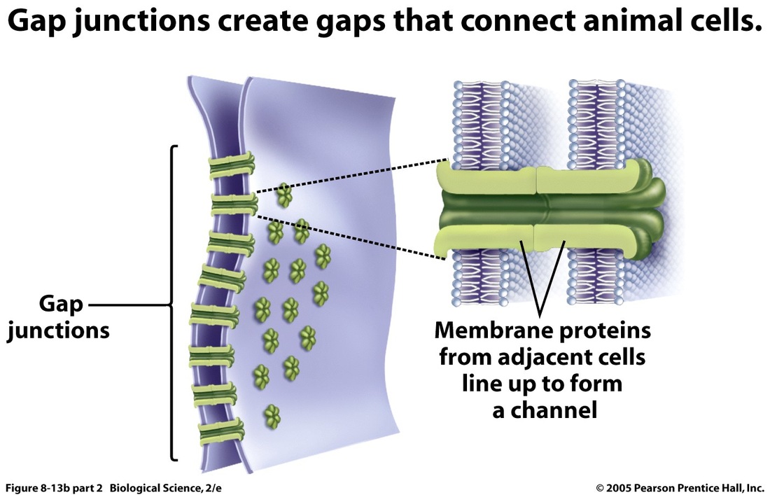
Gap junctions
Gap junctions serve the same function as and are considered analogous to plasmodesmata, but are found in animal cells as opposed to plant cells. Gap junctions allow nearly unlimited transportation of resources between adjacent cells, directly connecting the cytoplasm of neighboring cells. Just as with plasmodesmata, gap junctions can be turned "on or off" by opening and closing the connection and hence they require ATP to operate. As seen in the diagram to the right, there can be multiple gap junctions formed between two cells. |
|
Cellular Movement Organelles
|
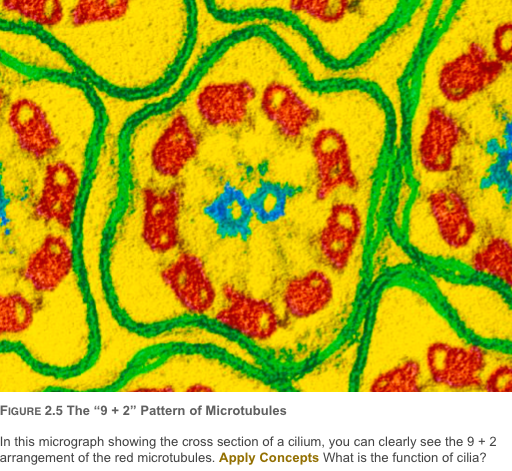
Flagella and cilia
Despite their names, cilia and flagella are actually the same organelle; flagella tend to be longer but cells have fewer of them, while cilia are more numerous, but shorter. Cilia and flagella are both composed of microtubules and help the cell to move. Some insect sperm cells have flagella than can reach 2 mm in length (that's enormous considering most cells are measured in micrometers). In mammals, epithelial cells are lined with cilia to help move debris and mucus and can reach astronomical numbers- more than 10^7/mm^2. Cilia and flagella are distinguished by their 9 +2 arrangement of microtubules: a pair of microtubules is surrounded by 9 microtubules. These structures enable cellular movement by beating or whipping, but this requires energy to do so. Cilia and flagella can be found in all bacteria and animal cells. |
|
Endosymbiotic Theory
|
Endosymbiotic Theory
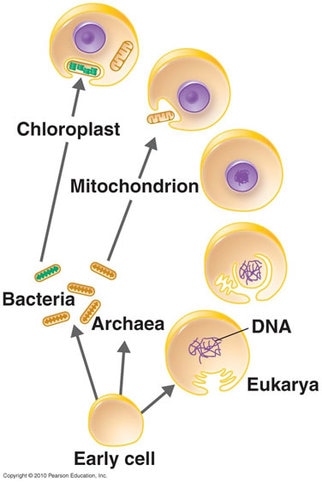
Chloroplasts and mitochondria are very unique from the other organelles, which has made biologists question their origin. Endosymbiotic theory explains the history of these two unique organelles. According to endosymbiosis, mitochondria and chloroplasts were once free-living, prokaryotic cells that were engulfed by larger, primitive eukaryotic cells. Normally the prokaryotic cells would have been digested for food, but the two cell types formed a symbiotic relationship and worked together to increase their survival. Over time, the prokaryotic cells surrendered their autonomy to become organelles, although have retained some their traits as free-living cells. |
|
How are Mitochondria and Chloroplasts Unique Organelles?
- Mitochondria and chloroplasts have their own DNA, unique from the cell. No other organelle has it own DNA as all cellular activity is controlled the by cell's DNA stored in the nucleus.
- Mitochondria and chloroplasts reproduce independently from the rest of the cell. While the other eukaryotic organelles do replicate themselves (to replace old or damaged organelles or to prepare for cell division), their replication is controlled by the cell or rather its DNA. Mitochondria and chloroplasts regulate their own replication which closely resembles the binary fission seen in bacteria cells.
- Mitochondria and chloroplasts have a double membrane. All other eukaryotic cells are membrane bound, but only in a single membrane, not two. The presence of an additional membrane around these two organelles highlights their uniqueness.
The Endosymbiotic Theory
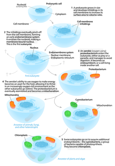
Millions of years ago, there were 3 basic types of primitive cells:
Millions of years ago, there were 3 basic types of primitive cells:
- Prokaryotic aerobic proteobacteria that were capable of cellular respiration. In cellular respiration, chemical energy in the form of glucose is converted into ATP as well has water and carbon dioxide. Cellular respiration can be aerobic or anaerobic menaing it can either use oxygen (aerobic) or not use oxygen (anaerobic). The aerobic proteobacteria used oxygen for cell respiration.
- Prokaryotic cyanobacteria capable of photosyntheis. Photosynthesis is the opposite chemical reaction of cellular respiration. In photoysntheis, carbon dixoide is combined with water and sunlight to form glucose and oxygen gas.
- Primitive eukaryotic cells that were much larger than the prokaryotic cells. These eukaryotic cells would regulalrly consume the smaller prokaryotic cells for food through the process of phagocytosis. Phagocytosis is a special type of endocytosis used to important bacteria cells into the cell.
|
The aerobic proteobacteria would eventually become the mitochondria in all eukaryotic cells, while the cyanobacteria would become chloroplasts seen in plant cells. The question is how? The aerobic proteobacteria and cyanobacteria were free-living cells complete with their own DNA, ability to reproduce (called binary fission) and a cell membrane. When these cells were engulfed by the eukaryotic cells through phagocytosis, they were encased in a vesicle which had the same composition as the cell membrane. This ultimately resulted in the bacteria cell (and the resuting organelles) having a double membrane. Rather than digesting and killing the bacteria cells (aerobic proteobacteria and cyanobacteria), the eukaryotic cell formed a mutualistic symbiotic relationship with them. The primitive eukaryotic cells (which would become modern plant, animal and fungal cells) were provided ATP by the aerobic proteobacteria and simple sugars by the photosynthetic cyanobacteria. In exchange the primitive mitochondria (aerobic proteobacteria) and chloroplasts (cyanobacteria) were offered a safe place to live. This explains why mitochondria and chloroplasts have their own DNA (a unique trait within organelles), have a double membrane and reproduce on their own- they were once free-living prokaryotic cells.
|
|
Why do Mitochondria and Chloroplasts have their unique traits?
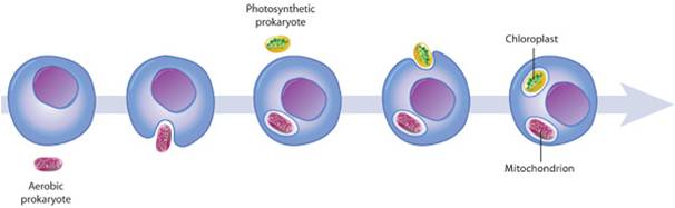
Because mitochondria and chlorplasts were once free living cells (aerobic proteobacteria and cyanobacteria respectively), they would have needed to fulfill all of the characteristics of life. This meant each cell had to have its own DNA and the ability to reproduce. Even after the aerboic proteobacteria and cyanobacteria were engulfed into the eukaryotic cell by phagocytosis and became the mitochondria and chloroplasts, they retained these traits. The double membrane can also be easily explained by the organelles' past life. As free living cells the aerboic proteobacteria and cyanobacteria would have had their own cell membranes, but when they entered the eukaryotic cell by phagocytosis, they were encased in another phospholipid bilayer as a vesicle formed around the prokaryotic cell. That vesicle would become the second membrane we see in both mitochondria and chloroplasts. This can be seen in the illustration below.
Because mitochondria and chlorplasts were once free living cells (aerobic proteobacteria and cyanobacteria respectively), they would have needed to fulfill all of the characteristics of life. This meant each cell had to have its own DNA and the ability to reproduce. Even after the aerboic proteobacteria and cyanobacteria were engulfed into the eukaryotic cell by phagocytosis and became the mitochondria and chloroplasts, they retained these traits. The double membrane can also be easily explained by the organelles' past life. As free living cells the aerboic proteobacteria and cyanobacteria would have had their own cell membranes, but when they entered the eukaryotic cell by phagocytosis, they were encased in another phospholipid bilayer as a vesicle formed around the prokaryotic cell. That vesicle would become the second membrane we see in both mitochondria and chloroplasts. This can be seen in the illustration below.
|
Which Came First: The Mitochondria or The Chlorplast?
We know mitochondria developed before chloroplasts because chloroplasts are only found in plant cells, while mitochondria are found in all eukaryotic cells including animal, plant and fungal cells. Therefore, mitochondria are an older "trait" and chloroplasts would have developed more recently. In other words, the traits that were present in the early eukaryotic cells (like mitochondria) would have been passed on to their offspring, while newer traits (like chloroplasts) would not be present in all the descendents of the primitive eukaryotic cells. |
|
|
The History of Other Eukaryotic Organelles
The endoplasmic membrane, Golgi apparatus and even the nucleus formed in a similar manner to the chlorplast and mitochondria. Although these organelles were never free-living cells, they did form because of infolding of the cell membrane. Early cells would often fold their membranes inward inorder to increase their surface area to volume ratio, allowing them to increase their nutrient exchange with the environment. Sometimes, these folds would "pinch off" completely, breaking away from the cell membrane and form internal membrane systems. These systems would go on to become the nucleus, ER and Golgi apparatus we know today. This is why those membraneous organelles share the same phopholipid bilayer sturucture with the cell membrane- they formed directly from the cell's plasma membrane. This process is similar to how the plasma membrane infolded on itself to engulf and import the cyanobacteria and aerobic proteobacteria via phagocytosis (this is the specific form of endocytosis that deals with bacteria cells). |
Bulk Transport: Endocytosis and Exocytosis
|
|
Vesicles: Endocytosis and Exocytosis
Sometimes the cell needs to move molecules that are simply too large or too numerous for protein channels/pumps, or even plasmodesmata/gap junctions to handle. This is when the cell relies on special processes called endocytosis and exocytosis. As the name suggests endocytosis (endo = into, cyto = cell, sis= process) is used to import large molecules or large quantities of molecules into the cell by infolding the cell membrane and engulfing the substance(s) in a vesicle. Both endocytosis and exocytosis are examples of active transport, meaning they require ATP to occur and are found in found primarily animal cells. Plant cells are unable to perform endo/exocytosis because of their cell wall. |
|
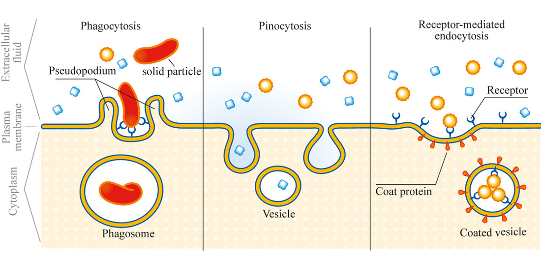
There are a number of different types of endocytosis including phagocytosis, pinocytosis and receptor-mediated endocytosis. Phagocytosis is often seen in immune cells and refers to the ingestion of bacteria cells (also known as phagocytes). The bacteria cell is surrounded by the cell's membrane and imported in a vesicle. Once the bacteria cells are ingested, they are destroyed by the immune cell. In a broader sense, phagocytosis can include the importation of any large substances into the cell.
|
|
This means that phagocytosis would also have been the specific type of endocytosis used by primitive eukaryotic cells to engulf cyanobacteria and proteobacteria according to the endosymbiotic theory. Pinocytosis refers to the ingestion of small particles by the cell, but unlike the other forms of endocytosis, no receptor proteins are used meaning a wide variety of substances can be imported. The particles gather on the outside surface of the cell membrane before the cell unfolds the membrane and captures the particles in a vesicle, thereby transporting them into the cell itself.
|
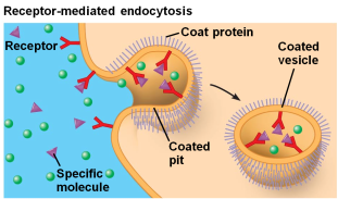
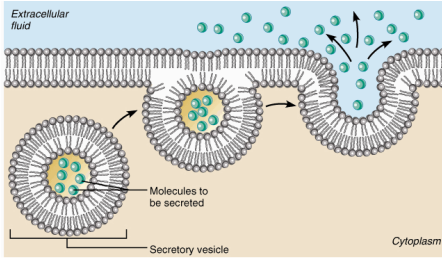
In receptor-mediated endocytosis, special receptor proteins embedded in the cell membrane bind with specific particles including other proteins, hormones and even some sneaky viruses. As with the other forms of endocytosis, the cell membrane folds inward surrounding and capturing the particles in a vesicle. However, receptor mediated endocytosis is regulated, meaning only specific particles can bind with the receptor proteins and thereby enter the cell. Exocytosis (pictured on the left) functions in a similar way, except the cell is expelling molecules. A vesicle within the cell will fuse with the cell membrane and force its contents out of the cell.
|
Cellular Transport
Passive and Active Transport
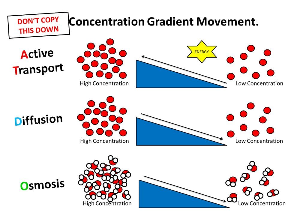
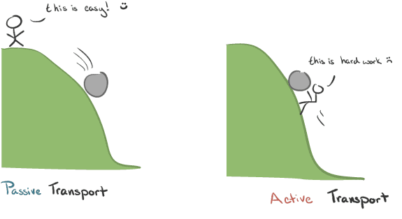
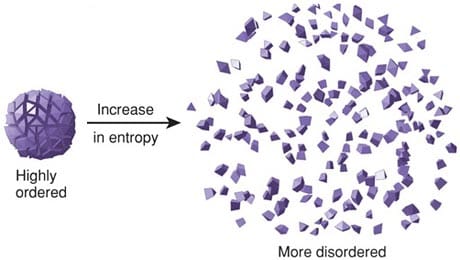
In general, resources can be moved around the cell (or between cells) in one of two basic ways: with energy or without energy. As the name suggests, passive transport does not require energy to occur and relies on natural processes. Passive transport relies on the principle of entropy; order cannot be maintained without energy and so systems move from order and organization to chaos and disorganization. In contrast, active transport does require energy to occur. While passive transport has a major advantage in that it does not require energy to occur and so the cell can basically use it for free, there are major drawbacks. Passive transport is usually slow and does not allow the cell to build a concentration gradient- it cannot build up a large concentration of the substance. This is because diffusion can only operate until equilibrium is achieved and all particles are evenly spread out. On the other hand, active transport requires energy (in the form of ATP) to operate, but is much faster than passive transport and allows the cell to build concentration gradients. So while, active transport is expensive for the cell, it is often more advantageous because of its speed and flexibility. We will discuss the various modes of passive and active transport below.
In general, resources can be moved around the cell (or between cells) in one of two basic ways: with energy or without energy. As the name suggests, passive transport does not require energy to occur and relies on natural processes. Passive transport relies on the principle of entropy; order cannot be maintained without energy and so systems move from order and organization to chaos and disorganization. In contrast, active transport does require energy to occur. While passive transport has a major advantage in that it does not require energy to occur and so the cell can basically use it for free, there are major drawbacks. Passive transport is usually slow and does not allow the cell to build a concentration gradient- it cannot build up a large concentration of the substance. This is because diffusion can only operate until equilibrium is achieved and all particles are evenly spread out. On the other hand, active transport requires energy (in the form of ATP) to operate, but is much faster than passive transport and allows the cell to build concentration gradients. So while, active transport is expensive for the cell, it is often more advantageous because of its speed and flexibility. We will discuss the various modes of passive and active transport below.
|
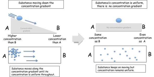
Concentration Gradient
A concentration gradient refers to the distribution of particles. The greater the difference in particle density between two areas, the greater the concentration gradient is said to be. You can think of the concentration gradient as a hill: particles will want to move down the hill, moving from high to low concentration. In this example, the particles move with the gradient, moving down the hill. As the particles move down the concentration gradient, the gradient becomes less and less steep until all particles are evenly distributed and equilibirum is achieved. In some cases, particles may move up the "hill", from low to high concentration, although this will require energy. In this context, the particles are said to move against the gradient, rather than with it. As the particles move against the concentration gradient, the gradient will become more and more steep as the particles become more and more concentrated. This is the exact opposite of the previous example. Passive transport moves particles along their concentration gradient (down the hill) from high to low concentration across the cell membrane without using energy by taking advantage of natural forces, but active transport moves particles against their gradient (up the hill) from low to high concentration across the cell membrane using energy. |
|
|
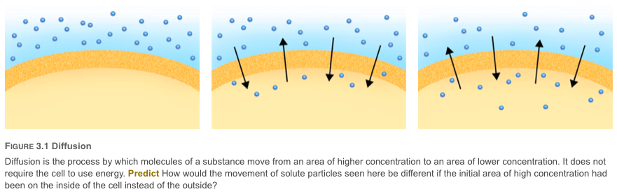
Diffusion
In general, particles move randomly: there is no pattern to their movement, except particles will move away from each other. Diffusion is the process by which particles move from areas of higher concentration (or density) to areas of lower concentration (or density). In other words, in diffusion particles move with their concentration gradient "down the hill." Diffusion is a form of passive transport and occurs as a result of a natural force called entropy. Entropy is the tendency of the universe to move from organization (high |
|
concentration) to chaos (low concentration). In other words, in the absence of energy entropy always increases much like how your room becomes increasingly messy (disorganized) over time unless you clean it (expend energy)
Here is an example of diffusion being driven by entropy: imagine placing a drop of red dye in a large beaker of water. At first the food dye particles will remain together, but over time the red particles of the dye will spread out within the water; in other words they diffuse. This is because of entropy: the particles in the drop are highly organized and begin to spread out in the beaker, becoming increasingly disorganzed.
Diffusion can be accelerated or catalyzed by supplying thermal energy. This is because heat is a measure of the average kinetic energy of the particles in a substance; adding thermal energy (heating up the substance) increases the kinetic energy of the particles allowing the particles to move more quickly. In other words, the warmer a substance is the faster diffusion can occur. The rate of diffusion is also influenced by the concentration gradient of the particles themselves. The steeper the gradient is, the faster diffusion will occur. Of course, as diffusion occurs, the less steep the gradient becomes, thereby slowing down the rate of diffusion. Diffusion will continue until equilibrium is achieved and all particles are spread out evenly. Therefore, you cannot concentrate particles (move them against their concentration gradient) using diffusion. In fact, if the concentration of particles outside the cell is lower than inside the cell, the particles will exit the cell as can happen with water and osmosis.
|
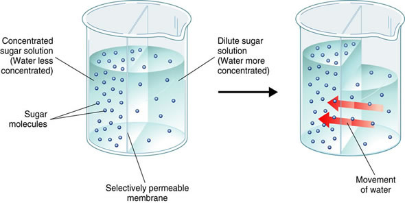
Osmosis
Osmosis is the diffusion of water and is therefore a type of passive transport. Osmosis follows all the rules of diffusion, water will always move toward areas of lower water concentration. Osmosis is particularly important to cells since all life requires water and plants use water to remain turgid and stand upright. As you know, water is regarded as the universal solvent and is capable of dissolving a wide variety of particles. The concentration of dissolved particles in water directly controls osmosis. When we are discussing osmosis, seven important terms need to be kept in mind and fully understood: |
|
- Solvent: a substance (generally a liquid, especially water) that is used to dissolve a solute
- Solute: particles that are dissolved into a solvent (usually water
- Solution: a homogenous mixture of 2 or more substances
- Tonicity: compares the relative concentration of solutes in 2 solutions. In this context, the solvent of the solution is always water. There are three types of tonicities that influence osmosis:
- Isotonic: both solutions have equal solute concentrations, no net water movement. Equilibrium has been achieved.
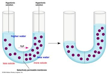
- Hypertonic: a hypertonic solution has a higher solute concentration than the other solution (which is hypotonic by definition) and therefore a lower water concentration. That may sound counterintuitive, but it makes sense: the higher the solute concentration you have in a solution, the lower the water concentration in that solution. Water will flow into the hypertonic solution until both solutions achieve equal concentrations of solute and are isotonic to each other. At this point, equilibrium is achieved.
- Hypotonic: a hypotonic solution has a lower solute concentration than the other solution (which is hypertonic by definition , which means the hypotonic solution has a higher water concentration. Again, this may sound counterintuitive, but it makes sense: the lower the solute concentration in a solution, the higher the water concentration must be. Water will flow out of the hypotonic solution until both solutions achieve equal concentrations of water and are isotonic to each other. At this point, equilibrium is achieved.
|
Osmosis Example
Imagine you had a cell with 15% solute concentration (by mass) suspended in a solution with 10% (by mass) solute concentration. If the cell is 15% solute, the remaining 85% is water. And if the solution surrounding the cell is 10% solute, the remaining 90% must be water. In this case, the cell is hypertonic and the surrounding environment of the cell is hypotonic. Water will rush into the cell, until equilibrium is achieved and both the cell and the environment have equal concentrations of solute and water. At this point the cell and the environment will be isotonic to each other. |
|
Cell Tonicity
When the cell has a higher concentration of solute (could be salt, sugar, etc.) than its surrounding environment, it conversely has a lower concentration of water within itself than its surrounding environment. This is because a solution (remember cells are aqueous and mostly water) is composed of only solute and solvent so as the concentration of solute increases, the concetration of solvent must decrease. In other words in a solution the concentration of solute and solvent are inversely proportional: as one increases the othe rmust decrease.
When the cell has a higher concentration of solute than its surrounding environment the cell is described as hypertonic and the surrounding environment is hypotonic. Water will rush into the cell as a result of osmosis (the water will move from high concentration --> low concentration) until equilibrium is achieved and the cell is isotonic relative to the surrounding environment.
Now imagine we reverse the previous situation: the concentration of solute within the cell is lower than the cell's surrounding environment and conversely, the cell has a higher concentration of water than the surrounding environment. Now the cell is hypotonic and the surrounding environment is hypertonic. Water will exit the cell via osmosis, effectively dehydrating the cell. This is what happens when you drink coffee, tea, soda or any other dehydrating beverage, the solute concentration within the drink is so high (and the concentration of water in the drink is so low) that it makes the surrounding environment of your cells hypertonic and draws water out of the cell. This continues until both the cell and its environment are isotonic and equilibrium is achieved.
Let's imagine one last situation: the concentration of solute is equal inside and outside the cell. In this case, the cell and the surrounding environment are both described as isotonic and there is no net movement of water into or out of the cell.
|
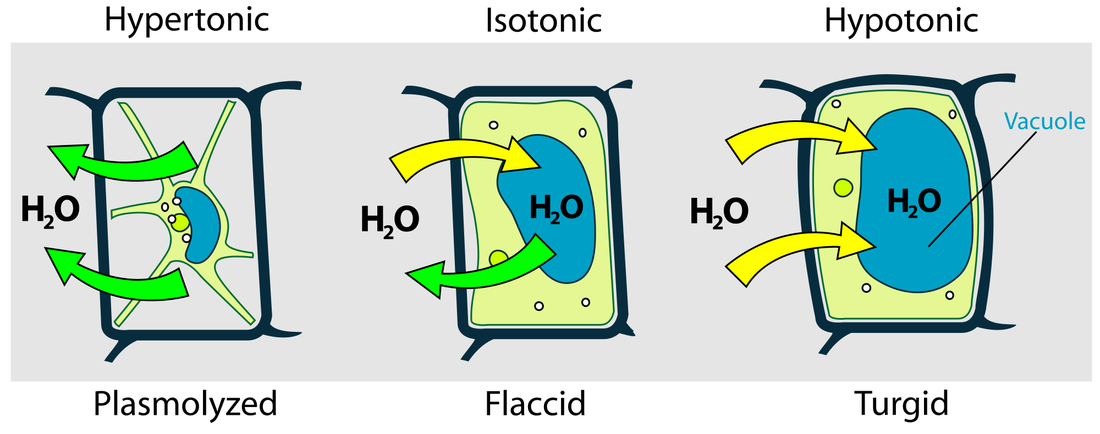
Different types of cells prefer different toncities. Consider plants, who rely on their large central vacuole to provide them with turgor pressure and help them stand up straight. It is very important that the plant cell remain hypertonic compared to its surrounding environment- this will force water into the cell and help the cell maintain turgor pressure. In fact, plants will actively pump salts and other solutes into their cells to keep up turgor pressure by forcing water to enter the cell via osmosis. Luckily the plant cell has a cell wall to keep it from bursting. Should the plant cell become isotonic the plant cell will become flaccid because the central vacuole will not be completely swollen with water. In a hypertonic environment, the plant cell can plasmolyze.
|
|
During plasmolysis, the central vacuole of a plant cell is emptied completely, eliminating all turgor pressure. As a result, the cell membrane collapses inward, shriveling up although still remianing attached to the cell wall in a few places. And finally, the cell wall itself will begin to pull inward taking a concave shape although largely remaning in tact.
In contrast to plants, animal cells generally require an isotonic environment. In a hypertonic solution, animal cells can shrivel up as water exists their cells via osmosis, although animal cells do not enter the same state of plasmolysis that plant cells do. In a hypotonic solution, water will rush into the animal cells which will swell in size. While this was a good thing for plant cells, it is actually very dangerous for animal cells since they lack cell walls. As the cell expands, it runs the risk of burst as a result of the increase in cell size due to osmosis, a process called lysis. Tonicity provides yet another reason for organisms to maintain homeostasis in their bodies.
In contrast to plants, animal cells generally require an isotonic environment. In a hypertonic solution, animal cells can shrivel up as water exists their cells via osmosis, although animal cells do not enter the same state of plasmolysis that plant cells do. In a hypotonic solution, water will rush into the animal cells which will swell in size. While this was a good thing for plant cells, it is actually very dangerous for animal cells since they lack cell walls. As the cell expands, it runs the risk of burst as a result of the increase in cell size due to osmosis, a process called lysis. Tonicity provides yet another reason for organisms to maintain homeostasis in their bodies.
Protein Channels and Aquaporins
We have already discussed these concepts before but now we can give them greater context. Because protein channels, like aquaporins, do not use energy they are involved in passive transport. Protein channels exist primarily to facilitate diffusion of materials that either cannot diffuse across the cell membrane, or do so too slowly. Consider water as an example. It is possible for water to move or diffuse across the cell membrane on its own, but water is polar and has difficulty moving through the phospholipid bilayer. Instead, aquaporin protein channels create special tunnels through the plasma membrane, enabling water to move into the cell with great easy. Aquaporins maintains the cell membrane's integrity by only allowing water molecules to pass through, other compounds are excluded. Remember the primary goal of the cell membrane is to regulate what enters and exits the cell. In short, protein channels like aquaporins allow for facilitated diffusion a type of passive transport.
We have already discussed these concepts before but now we can give them greater context. Because protein channels, like aquaporins, do not use energy they are involved in passive transport. Protein channels exist primarily to facilitate diffusion of materials that either cannot diffuse across the cell membrane, or do so too slowly. Consider water as an example. It is possible for water to move or diffuse across the cell membrane on its own, but water is polar and has difficulty moving through the phospholipid bilayer. Instead, aquaporin protein channels create special tunnels through the plasma membrane, enabling water to move into the cell with great easy. Aquaporins maintains the cell membrane's integrity by only allowing water molecules to pass through, other compounds are excluded. Remember the primary goal of the cell membrane is to regulate what enters and exits the cell. In short, protein channels like aquaporins allow for facilitated diffusion a type of passive transport.
|
Protein Pumps and Endo/Exocytosis
Again, we have already discussed these concepts earlier, but now we can give them greater context. Sometimes the cell needs to build up large supplies of a substance or needed to move particularly large molecules or maybe a large quantity of molecules. In these situations, diffusion (even facilitated) is not going to cut it. Protein pumps like the potassium chloride pump allow cells to move huge quantities of potassium ion into the cell, against the potassium ion's concentration gradient (low --> high concentration, the opposite of diffusion). But protein pumps require energy and hence fall under the category of active transport. When the cell needs to move large molecules like glucose or large numbers of molecules into the cell, it uses endocytosis (endo = into, cyto = cell). The molecules being imported bind to receptors on the outside of the cell membrane, regulating what enters the cell. The cell membrane folds inward and pinches off, forming a vesicle around the molecules which have been successfully imported into the cell. This requires energy (ATP) and hence fits into the category of active transport. The reverse of endocytosis is exocytosis, where the molecules are exported out of the cell. A vesicle, filled with the molecules to be removed the cell, travels to the plasma membrane and merges with it. The molecules spill out of the cell and are successfully exported from the cell. |
|
Cellular Differentiation
As you know, the cell is the most basic unit of life: cells are the smallest unit of structure and function in an organism that still retain the characterisitics of life. Smaller units of structure such as organelles, molecules and atoms are not considered alive despite being relevant to higher orders of structure like cells. In single celled organisms including most prokaryotes, that one cell is responsible for carrying out all of the characterisitics of life such as maintaining homeostasis. However, in multicellular organisms such as most eukaryotes, cells can divide up their responsibilties and work together. This leads to cell differentiation in which cells become specialized in their function. Each cell will take on a specific role designed to help support the organism as whole. For example, some cells will become bone cells, other blood cells and still others will become nerve cells.
|
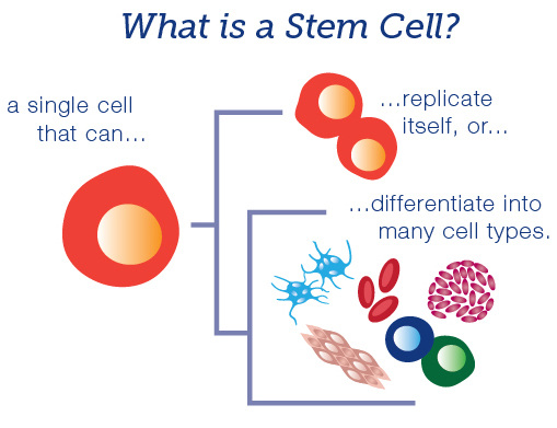
Stem Cells
Cells that are undifferentiated are known as stem cells, which can self-replicated and develop into essentially any type of cell in a multicellular organism. Stem cells can be thoughout of as the stem of a plant that branches out into all the different cell types. There are three major groups of stem cells: totipotent, pluripotent and multipotent. Totipotent stem cells (fetal stem cells) are the stem cells present after the zygote undergoes its first few round of cell division. These cells can form whole new organisms.Pluripotent stem cells (embryonic stem cells) quickly replace totipotent cells and can become any type of cell in the organism without restriction and are found in embryos, but unlike totipotent cells, are incapable of forming a completely new organism. These pluripotent cells help the embryo to develop, but become less |
|
prevalent as the organism changes, disppaearing entirely by adulthood. Multipoint stem cells (adult stem cells) have the ability to become more than one type of cell but are more limited than pluirpotent cells. Unlike, pluripotent cells, multipotent cells persist in adult organisms and are found in bone marrow and the brain as adult stem cells, replacing dead or diseased cells.
|
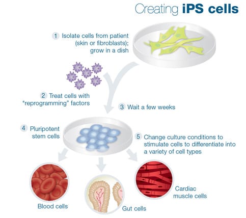
Induced pluripotent stem cells (iPSCs) are adult stem cells (multipotent) that have been reverted (induced) into embryonic stem cells (pluripotent) using a variety of chemical factors. iPSCs are useful because they allow for scientific research on and medicial treatment with embryonic stem cells without the need to harvest and destroy embryos. Adult stem cells can be harvested without killing the organism and help ensure that the patient's body will not reject the stem cells as part of a medical treatment since they are in fact the patient's on cells.
Currently, the major drawbacks of iPSCs are that these cells are slow to cultivate and the process can be expensive to perform. But, the biggest challenge is how to maintain the cells, storing them for future research and use. However, given the inherent benefits of using iPSCs, it is likely that the drawbacks will become less prevalent as better technologies and cultivation techniques become available. |
Induced pluripotent cells were first produced in 2006 by Shinya Yamanaka’s team in Kyoto, Japan. Yamanaka won a Nobel Peace Prize in 2012 for his work.
|
Levels of Organization
In multicellular organisms, cells work together to support the survival of the organism as a whole. This creates specialized levels of organization with in the organism including: cells, tissues, organs and organ systems. Groups of cells with the same function (ie. bone cells or blood cells) combine to form tissues which in turn can combine to form organs. Organs are self contained parts of an organism that have specific, vital roles to the organism. Organs with similar functions are generally grouped together to form organ systems, working together to perform a specific function. In this way, each cell is assigned a specific function or role and works together with other, similar cells to perform a function that supports the organism as a whole. |
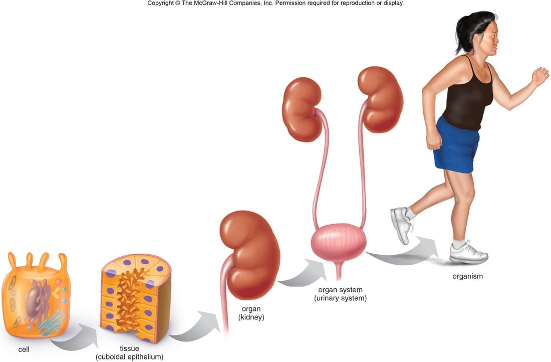
|
For example, a group of cardiac cells would form cardiac tissue which in turn forms your heart (an organ). Your heart along with your blood vessels (including arteries, veins and capillaries) would make up your cardiovascular system, one of the major organ systems in your body.
